


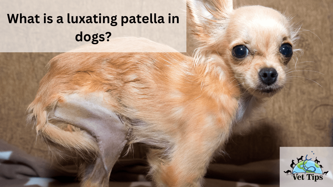
Here in this article, we discuss all about, “What is a luxating patella in dogs?”. Continue reading to learn more about it.
The dislocation of the patella bone is known as patella luxation. The patella runs up and down in a groove on the thigh bone. A luxating patella can move around in the patella groove, slipping in and out of its usual location. Patella luxation is a widespread problem in small young dogs like Pomeranians, Poodles, and Shih Tzus, and it can also happen in large breed dogs.
What causes luxation of the patella?
It is a congenital problem caused by the patella tendon’s irregular orientation, which is usually towards the inside of the leg. As the dog grows older, the issue will become more severe.
Patients with PL may have varying degrees of lameness on an affected limb, ranging from asymptomatic to non–weight-bearing. The severity of the luxation, the disorder’s progression and later joint capsule changes, exercise duration, and orthopedic disruptions all affect clinical signs.
Anatomy (Form and Structure)
The patella is known as a sesamoid bone encased in the patellar ligament. It is located at the distal end of the quadriceps femoris muscle that connects to the tibial tuberosity. The four muscle heads join to form a single quadriceps femoris muscle.
The patella is a stable bone and functions as a pulley in a dog’s body. Normal patellar axial alignment allows the patella to exert adequate pressure on the trochlear groove’s articular cartilage during neonatal and adolescent development, resulting in a groove with sufficient width and optimal depth as the dog develops.
Trauma or congenital disabilities can cause patella luxation; congenital disabilities are much more common. Congenital luxations are more common in dogs of any weight, age, or species; according to a 1994 survey, 82 percent of the study’s dogs had congenital luxations. The breeding of affected congenital patients leads to its increased number of cases nowadays.
Multiple anatomic disabilities can cause PL. Bowing of torsion of the tibia leading to tibial deformity, epiphyseal dysplasia of femur, displacement/atrophy of the big muscle, i.e., quadriceps femoris, hip dysplasia, deep trochlear groove with missing or hypoplastic trochlear ridges, and rotational deformation of the stifle joint are all potential underlying musculoskeletal defects that may lead to patellar luxation in canines.
Patellar Luxation Grades (What is a luxating patella in dogs?)
First grade
When the limb is extended, the patella may be manually luxated, but it returns to the trochlear groove as soon as pressure is elevated. The joint should be able to stretch and expand usually. During natural joint motion, spontaneous luxation of the patella is unusual.
Second grade
The patella may be luxated manually or naturally when the stifle is flexed. The patella stays relaxed until it returns to the trochlear groove spontaneously with energetic stifle extension or until it is removed by hand/manually.
3rd grade
The patella is continually flexed/luxated, but it is quickly replaced by side. When manual pressure is withdrawn from the patella, it reluxates spontaneously.
4th grade
The patella is luxated all of the time and cannot be decreased manually.
CLINICAL SIGNS
To distinguish the severity of clinical signs and symptoms present during physical examination, Patellar Luxation is rated on a scale of 1 to 4. Grade 1 Patellar Luxation is often discovered by accident during routine exams, with the affected dog presenting asymptomatically and the pet parents stating no clinical indication. Dogs with grade 4 or 3 Patellar Luxation usually have consistent or intermittent hindlimb lameness and are hesitant to leap or walk down a hill.
Owners may have heard and felt a “click” or “pop” when walking their dog or found that the dog would abruptly kick it out to the caudally or side. Sometimes, dogs stop and look back toward the affected limb or decrease the luxation without further interference. Patients with grade 4 luxation are often lame or non–weight-bearing on the affected leg, with weight moved cranially during stance and walking. During ambulation, they often are unable to stretch the affected stifle fully.
Patients’ ages at which it shows signs and symptoms usually vary but generally, it is ranged from 2 to 6 years in the case of dogs.
If the bodyweight is increased, articular cartilage damage and a concurrent cranial cruciate ligament (CCL) rupture may occur, which can worsen this condition. Affected dogs have a crouching or shuffling gait and exhibit occasional locking of the stifle after extension. Lameness is not necessarily present in affected small dogs or cats.
DISEASE DIAGNOSTIC
The most common way to diagnose PL is to palpate the affected stifle. Radiography may be used to assess limb deformities’ presence and nature and schedule any surgical realignment procedures. If osteoarthritis is present, radiography may also be used to determine the severity of the condition. In grade 3 and 4 PL, the patella will be visible outside of the radiography trochlear sulcus. In contrast, in grade 1 and 2 luxations, the patella may be visible inside the trochlear sulcus.
OPPORTUNITY
In dogs and cats, medial patellar luxation (MPL) is much more prevalent and dangerous than lateral patellar luxation (LPL), regardless of the patient’s age or size. According to research, MPL affects 83 percent to 96 percent of all canines diagnosed with Patellar Luxation. In contrast, LPL affects big- and giant-breed dogs more than tiny or toy-breed dogs, with a prevalence of up to 33 percent in giant breeds.
Miniature or Toy breed of dogs, such as Chihuahuas, Cavoodles, Pekingese, miniature poodles, Pomeranians, Maltese, and Yorkshire terriers, well as mixed breeds and Labrador retrievers, Flat retrievers are the most often diagnosed with PL.
The evidence for sex preference is mixed. According to some research, male-to-female ratios vary from 1:1 to 1.7:1 to 1:1.8. According to one hypothesis, male dogs large-breed and female dogs small-breed are more likely to have PL.
On average, 50 percent of dogs diagnosed with MPL having bilateral luxations (range: 41 percent to 83 percent). Bilateral PL was observed in 38 percent of cats in one sample and 81 percent of cats in the other. Concurrent CCL has been discussed and studied in patients with PL, with rates ranging from 4% to 41%.
According to most experts, dogs with bilateral Medial Patella Luxation had an advanced grade of stifle luxation and concomitant CCL rupture than canines with lower patellar luxation grades. It means that dogs with grade 4 Medial Patella Luxation were substantially more likely to have concomitant CCLR than dogs with all other luxation grades.
TREATMENT OF PATELLAR LUXATION
Surgical procedures (What is a luxating patella in dogs?)
Treatment of Patellar Luxation depends on the type and nature of the luxation, coexisting conditions with PL, the dog’s age, and any personal or economic restrictions. Surgery is usually the best treatment for grade 3 or 4 luxation; however, opinions differ among vet-docs, even among professional surgeons, when recommending surgery for grade 2 PL.
Some surgeons recommend surgery for dogs with grade 2 luxations with mild, acute osteoarthritis with intermediate lameness, while others recommend only conservative care. Surgical treatment of PL is often prescribed for puppies and young adult dogs to avoid immature articular cartilage wear in the nearby joints, such as the stifle joint. Surgery is advised in patients with active growing phases to prevent skeletal deformities from worsening.
Medial Patellar Luxation Surgical Interventions
1. Reconstruction of the bones
Trochleoplasty is a procedure that involves the removal of a piece of cartilage from a trochlear groove.
- Recession of the trochlear wedge completely
- Recession of the immediate trochlear block
- Trochlear septoplasty
- Trochlear chondroplasty in puppies under six months of age
- Wedge trochleoplasty of the medial ridge
- Transposition of the tibial tuberosity
2. Restoration of soft tissues
- Retinal tissues are released
- Desmotomy is the first step in the process.
- Capsulectomy (Removal of the capsule)
- Imbrication of the retinaculum
- Removal of obsolete retinal tissue
- Sutures that prevent rotation
- Quadricep muscles contraction
The purpose of this surgery is to position the quadriceps extensor muscle, reestablishing normal joint function by fixing the patella bone in the trochlear groove of the femur. This can help to slow the spread of underlying musculoskeletal diseases like osteoarthritis. Most sources agree that Patella Luxation surgery should provide many surgical correction techniques to optimize the effective return of function.
These procedures can be classified into two major categories, i.e., bone reconstruction and soft tissue reconstruction. Dogs with grade 4 luxations would almost certainly need multiple corrective tissue surgeries, and pet parents should know this possibility. Restricted movement and supervised physical therapy activities are part of postoperative treatment to facilitate recovery and avoid infection or complications. Before the sick dog is allowed to participate in an elevated activity after a tibial tuberosity transposition, radiography should be conducted at 7 to 8 weeks to determine proper bone healing.
POC and after surgical management. (What is a luxating patella in dogs?)
Pain relief with potent analgesics and (NSAID) nonsteroidal anti-inflammatory medications and various nutraceuticals recommended by the veterinarian are typical conservative treatments for PL.
Application of cold therapy/massage to the stifle joint area provides an analgesic effect, minimizes inflammation, and is made at home by pet parents appropriately trained. Acupuncture (TENS) Transcutaneous electric nerve stimulation, laser treatment, and clinical X-rays and ultrasound are examples of methods to cure dogs and relieve pain.
The following exercises will help you improve your dog hindlimbs in general after surgery:
- Inclined stand
- 3-legged or 2-legged stands
- Sit & stand(half squat)
- Sidestepping
- Walking uphill
- Running on an underwater treadmill with water levels ranging from stifle joint to mid-thigh region
- Full-range sit to stand
What if my pet isn’t a candidate for surgery?
That is purely determined by your vet depending upon your dog’s condition and PL grade. It is advised to consult at least 2 or 3 vet docs before going for surgery in case of PL.
Tell us in comments how you like our article: What is a luxating patella in dogs?
For similar posts click here.
For the source file click here.


One thought on “What is a luxating patella in dogs?”